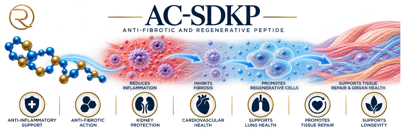
What the studies actually found
After a simulated heart attack, treated rats saw heart collagen (scar tissue) drop from roughly 23 to 15 micrograms per milligram of tissue, and inflammatory immune cells fall by about 36% — whether the treatment started before or after damage had already set in.
READ THE RESEARCH →Ac-SDKP is normally broken down by ACE, the same enzyme that common ACE-inhibitor blood pressure medications block. Human studies confirm that taking an ACE inhibitor raises circulating Ac-SDKP roughly 4- to 5-fold — a real, measured human effect, not just an animal finding.
READ THE RESEARCH →Ac-SDKP was first known for slowing cell division. Researchers were surprised to find it also drives new blood-vessel formation in living tissue, with effects on vessel growth comparable to a known angiogenic growth factor.
READ THE RESEARCH →Ac-SDKP's entire research story started in hematology: it was isolated as a natural regulator that protects blood-forming stem cells from the toxicity of chemotherapy and radiation. Its antifibrotic effects weren't discovered until years later.
READ THE RESEARCH →Ac-SDKP is the exact N-terminal 4 amino acids of Thymosin Beta-4 — the same parent molecule behind TB-500. One enzyme cut is all it takes to turn one into the other, yet the two go on to do very different jobs in the body.
EXPLORE THE SCIENCE →Ac-SDKP — Overview
What Is Ac-SDKP?
Ac-SDKP, formally N-acetyl-seryl-aspartyl-lysyl-proline, is a naturally occurring four-amino-acid peptide (tetrapeptide) found in the body — also known as goralatide and seraspenide. Unlike many research peptides engineered from naturally occurring hormones, Ac-SDKP is itself a physiological molecule the body produces as part of normal peptide processing.
Its scientific story has changed considerably over time. It was first identified for its ability to regulate hematopoietic stem and progenitor cells — the cells responsible for producing blood cells. Decades later, researchers recognized a second important characteristic: Ac-SDKP appears to act as a natural counterweight to fibrosis, the excessive accumulation and remodeling of connective tissue that can progressively impair organ structure and function. That shift — from a peptide associated primarily with bone-marrow biology to one investigated across cardiac, renal, pulmonary, vascular, and inflammatory research — is one of the most interesting aspects of Ac-SDKP (the full arc is on the Research page).
Ac-SDKP is also closely connected to thymosin beta-4 (Tβ4): its four amino acids form the N-terminal sequence of Tβ4, a 43-amino-acid peptide involved in several aspects of cellular biology, and research indicates Ac-SDKP can be generated from Tβ4 through enzymatic processing involving meprin-α and prolyl oligopeptidase. That relationship gives Ac-SDKP an unusual biological identity — it is both an independent signaling peptide and a small product generated during processing of a larger endogenous peptide.
Scientific Discovery & Development
The story began in 1989, when Maryse Lenfant, Joanna Wdzieczak-Bakala, and colleagues reported isolating and structurally characterizing a previously unknown tetrapeptide from fetal calf bone marrow.¹ They identified Ac-Ser-Asp-Lys-Pro and found it strongly inhibited proliferation of pluripotent hematopoietic stem cells — raising the question of why bone marrow would contain a small peptide capable of keeping stem cells relatively quiescent rather than continuously proliferating. Subsequent work in the early 1990s established Ac-SDKP as a physiological regulator of hematopoietic stem-cell activity, produced under normal conditions and present in biological tissues and fluids. At this stage, it was viewed primarily through the lens of blood-cell production and stem-cell regulation.
The story expanded when investigators found Ac-SDKP was connected to thymosin beta-4: in 1990, Grillon and colleagues showed Tβ4 could serve as its precursor, with prolyl oligopeptidase later identified as an important enzyme in the generation pathway.
Another major discovery came from angiotensin-converting enzyme (ACE) research: ACE was found to be responsible for degrading Ac-SDKP. This mattered because ACE inhibitors — widely used cardiovascular medications — reduce Ac-SDKP breakdown and consequently raise its circulating levels; human studies of ACE inhibition have shown substantial increases in plasma Ac-SDKP.² This changed the scientific question again: could some of the tissue-protective effects historically associated with ACE inhibition involve Ac-SDKP itself, rather than being explained entirely by changes in angiotensin II? That question moved Ac-SDKP into cardiovascular and fibrosis research.
Why Researchers Are Studying Ac-SDKP
The central interest today is its apparent relationship with tissue remodeling, inflammation, and fibrosis. Fibrosis isn’t simply “scar tissue” — it’s a repair response that becomes damaging when it stays active too long: fibroblasts become highly active, extracellular matrix proteins like collagen accumulate, and normal tissue architecture gradually distorts. This can occur in the heart, kidneys, lungs, liver, and other organs, which is why researchers are interested in molecules that help preserve the balance between repair and excessive fibrosis.
Ac-SDKP has emerged as one such candidate: experimental research has repeatedly associated it with reduced collagen accumulation, decreased fibroblast activity, modulation of inflammatory responses, and attenuation of fibrotic remodeling in several animal and cellular models. Importantly, this does not mean Ac-SDKP has been demonstrated to treat fibrosis in humans — most of the therapeutic evidence remains preclinical (see Research). Its significance lies in the biological pattern researchers have observed and the possibility that this naturally occurring peptide is part of the body’s own system for regulating excessive tissue remodeling.
Biological Foundation
Ac-SDKP is best understood as part of a larger biological conversation between injury, inflammation, repair, and remodeling. When tissue is damaged, the body activates inflammatory and repair programs — cells recruit immune cells, stimulate fibroblasts, modify the extracellular matrix, and rebuild tissue. One of the most important pathways in fibrotic remodeling is transforming growth factor-beta (TGF-β): essential for normal repair, but capable of pushing fibroblasts toward excessive extracellular-matrix production when signaling is prolonged or excessive. Research suggests Ac-SDKP can influence aspects of this profibrotic environment — studies in cardiac, renal, and pulmonary models have linked it with reduced collagen deposition, and cellular studies have shown inhibition of TGF-β-associated fibrotic responses (mechanistic detail is on the Science page).
Ac-SDKP is also unusual because no specific dedicated receptor has been definitively established as the explanation for its effects — researchers have instead investigated its interactions with broader signaling networks and its relationship with enzymes like ACE and prolyl oligopeptidase. That makes it less like a conventional hormone acting through one clearly defined receptor and more like a small regulatory component embedded within several interconnected biological systems.
What Researchers Are Investigating
Kidney — whether Ac-SDKP can reduce collagen and fibronectin accumulation, limit activated myofibroblasts, and suppress other features of progressive renal fibrosis; animal models of kidney injury have shown reduced fibrotic remodeling with treatment.
Lung — in a mouse model of bleomycin-induced pulmonary fibrosis, Ac-SDKP administration was associated with reduced inflammation, lung injury, collagen accumulation, and fibrosis; research using human lung fibroblasts has reported inhibition of TGF-β-induced myofibroblast formation and collagen production.
Cardiovascular — models of cardiac fibrosis and tissue remodeling helped establish the broader concept that Ac-SDKP may act as an endogenous antifibrotic factor beyond its original hematopoietic role.
ACE inhibitors — because ACE breaks down Ac-SDKP, ACE inhibition causes it to accumulate, leading researchers to investigate whether increased Ac-SDKP contributes to some of the tissue-protective effects seen with ACE inhibition. The relationship is biologically compelling, though it doesn’t establish that Ac-SDKP is solely responsible for ACE inhibitors’ clinical effects.
Stability and analogues — because the natural peptide is susceptible to enzymatic degradation, researchers are studying degradation-resistant analogues as tools for understanding — or eventually extending — its biological activity.
Potential Significance
The significance of Ac-SDKP comes less from a single proposed “benefit” and more from the possibility that it helps regulate how tissues respond to injury: Ac-SDKP → modulation of inflammatory and fibrotic signaling → reduced excessive extracellular-matrix accumulation → preservation of tissue architecture. In experimental models this pattern has appeared in several organs, most strongly in preclinical cardiac, kidney, and pulmonary fibrosis models — making Ac-SDKP an interesting candidate for studying whether endogenous antifibrotic mechanisms can be harnessed therapeutically. Its original connection to hematopoietic stem-cell regulation adds another dimension, with effects on inflammation, vascular biology, and cellular survival that remain active areas of investigation. The important distinction is between biological potential and demonstrated clinical benefit — the experimental evidence shouldn’t be read as proof the peptide produces the same effects in humans.
Current Research Stage & Future Outlook
Ac-SDKP remains investigational and is not FDA approved as a therapeutic drug. Its research history is unusual in that the molecule has been studied in humans in limited contexts — including its relationship with ACE inhibition and investigations involving an Ac-SDKP-related compound for renal physiological measurements — but these studies don’t establish an approved antifibrotic therapy, and there is currently no established Phase 1–3 clinical program demonstrating Ac-SDKP as an effective treatment for organ fibrosis.
The major scientific challenge is translation: animal studies have produced compelling findings across several organ systems, but researchers still need to determine whether those effects reproduce safely and consistently in humans, and to understand pharmacokinetics, optimal exposure, degradation, biological targets, and whether more stable analogues offer advantages over the natural molecule. That gap between strong biological rationale and limited human therapeutic evidence is what makes Ac-SDKP an important research subject rather than an established treatment.
Global Research Perspective
Ac-SDKP research spans an international network across hematology, cardiovascular biology, nephrology, pulmonary research, pharmacology, and regenerative biology. The original discovery came from European research into hematopoietic stem-cell regulation; subsequent work on ACE, cardiovascular remodeling, kidney fibrosis, and pulmonary fibrosis has involved researchers across Europe, North America, Asia, and other regions — including Maryse Lenfant, Joanna Wdzieczak-Bakala, Maria Cavasin, Oscar Carretero, Nitin Kumar, and many collaborators who helped establish the peptide’s biological story. The field has evolved through a sequence of discoveries rather than one breakthrough: first a hematopoietic regulator, then a thymosin beta-4-derived peptide, then an ACE substrate, and finally a candidate endogenous regulator of fibrosis and tissue remodeling.
Why This Matters
Ac-SDKP is scientifically interesting because its story reaches far beyond its four amino acids — from a bone-marrow discovery, to a link with thymosin beta-4 and ACE, to a potential endogenous regulator of inflammation and fibrosis across the heart, kidneys, lungs, and other tissues. The most important open question isn’t whether Ac-SDKP produces intriguing effects in experimental systems — it clearly does — but whether those effects can translate into meaningful human applications. That distinction defines the next chapter. The Research page covers how scientists arrived at the current evidence, while the Science page goes deeper into the molecular pathways, enzymatic processing, signaling networks, and pharmacology that make Ac-SDKP such an unusual peptide.
References
- Lenfant M, Wdzieczak-Bakala J, et al. Isolation and structural characterization of a tetrapeptide inhibitor of hematopoietic stem cell proliferation from fetal calf bone marrow. Discovery/mechanism literature — original and related papers include the tetrapeptide’s mechanism-of-action studies: https://pubmed.ncbi.nlm.nih.gov/8241953/ and https://pubmed.ncbi.nlm.nih.gov/1457609/
- Azizi M, et al. Acute angiotensin-converting enzyme inhibition increases the plasma level of the natural stem cell regulator N-acetyl-seryl-aspartyl-lysyl-proline. Journal of Clinical Investigation. 1996. https://www.jci.org/articles/view/118484
Ac-SDKP — The Research
Ac-SDKP has one of the more unusual research histories among short bioactive peptides. It wasn’t originally pursued as a modern therapeutic peptide — scientists first encountered it as a naturally occurring regulator of blood-forming stem cells. Only later did research reveal the same small molecule was deeply connected to fibrosis, inflammation, angiogenesis, tissue injury, and the angiotensin-converting enzyme (ACE) system. That shift — from hematopoietic regulator to endogenous antifibrotic signaling molecule — is the central story of Ac-SDKP research.
How Was Ac-SDKP Discovered?
The earliest clue came from hematology research in France. Researchers at the Institut Gustave-Roussy and French INSERM laboratories were studying why some bone-marrow stem cells remain dormant rather than continuously entering the cell cycle. A small inhibitory activity was identified in bone-marrow extracts, eventually leading to isolation of the tetrapeptide now called N-acetyl-seryl-aspartyl-lysyl-proline. By 1989, researchers including J. Wdzieczak-Bakala, M. Lenfant, and Eric Frindel had established that Ac-SDKP could suppress entry of primitive hematopoietic cells into DNA synthesis; mouse experiments suggested it helped maintain these cells in a quiescent state, and neutralizing endogenous Ac-SDKP caused substantially more stem cells to enter the cell cycle.¹
That raised the next question: where was this peptide coming from? Around 1990–1991, researchers demonstrated that Ac-SDKP could be generated from thymosin β4, a naturally occurring 43-amino-acid peptide — Ac-SDKP corresponds to Tβ4’s N-terminal sequence, produced through enzymatic processing. This connected a previously isolated bone-marrow regulator to a much larger endogenous peptide-processing system.
The next breakthrough came from ACE. Researchers found ACE does more than participate in the renin-angiotensin system — it also degrades Ac-SDKP. In 1996, Michel Azizi and colleagues at Hôpital Broussais in Paris demonstrated this directly in humans: in a double-blind crossover study of eight healthy volunteers, a single 50-mg dose of captopril produced a 5.5-fold increase in plasma Ac-SDKP while strongly inhibiting its breakdown.² This changed the scientific question again — Ac-SDKP was no longer simply a hematopoietic peptide, but appeared to be part of a broader physiological system connecting peptide production, ACE activity, stem-cell regulation, and tissue biology.
The Research Moves Toward Fibrosis
During the 1990s and early 2000s, researchers began asking whether Ac-SDKP influenced other rapidly remodeling tissues. Work from the cardiovascular research group led by Oscar A. Carretero at Henry Ford Hospital in Detroit showed Ac-SDKP could inhibit fibroblast activity and reduce pathological collagen accumulation. A landmark 2003 Hypertension study by Hongmei Peng and colleagues asked a sharper question: could Ac-SDKP merely prevent fibrosis, or actually reverse fibrosis already established? Using hypertensive rats with existing cardiac fibrosis, researchers administered Ac-SDKP for eight weeks; the peptide reduced left-ventricular collagen in a dose-dependent manner, and importantly, the intervention began after hypertension and fibrosis were already established.³ That distinction mattered — the research was moving from a prevention model toward the possibility of actively remodeling established fibrotic tissue, and the findings connected Ac-SDKP to the TGF-β pathway, one of the major systems driving pathological fibrosis.
A Second Surprise: Ac-SDKP Promotes Angiogenesis
The research became more interesting because Ac-SDKP didn’t simply suppress cell activity. In 2004, Dahai Wang, Oscar Carretero, Xiao-Ping Yang, and colleagues at Henry Ford Hospital reported that Ac-SDKP stimulated endothelial-cell proliferation, migration, and tube formation — in animal models, it increased new blood-vessel formation and myocardial capillary density after infarction.⁴ This created an unusual biological profile: Ac-SDKP appeared able to restrain excessive fibroblast activity while supporting endothelial responses associated with new vessel formation — a distinction that mattered in later research, since successful tissue repair needs more than suppressed fibrosis; it also needs blood supply, controlled inflammation, and remodeling.
Research Findings by Major Biological Area
Cardiac fibrosis and remodeling. The heart became one of the strongest areas of Ac-SDKP research. Animal studies repeatedly found reduced cardiac collagen deposition and fibrosis in models of hypertension, diabetes, myocardial infarction, and other cardiac injury. A 2010 study exposed human cardiac fibroblasts to TGF-β1, a powerful profibrotic signal; Ac-SDKP suppressed the transformation of fibroblasts into myofibroblasts, reduced collagen production, and inhibited Smad2 and ERK1/2 signaling.⁵ The significance: this antifibrotic effect wasn’t just an observation in an animal heart — Ac-SDKP could directly influence a human cardiac cell type involved in extracellular-matrix production. A more recent 2024 study from researchers associated with Henry Ford and Wayne State University found Ac-SDKP also reduced collagen production in human cardiac fibroblasts exposed to endoplasmic-reticulum stress, linking the effect to CHOP/NF-κB inflammatory signaling.⁶ The cardiac research has progressively moved from an observation (“Ac-SDKP reduces fibrosis”) toward a detailed picture of the cellular processes it can influence in pathological remodeling.
Kidney. In 2013, Yiqin Zuo, Agnes Fogo, Nour-Eddine Rhaleb, Oscar Carretero, and collaborators at Vanderbilt and other institutions investigated Ac-SDKP in renal fibrosis models: it reduced collagen and fibronectin deposition, decreased myofibroblast and macrophage accumulation, and suppressed profibrotic factors.⁷ A separate study in diabetic rats found Ac-SDKP reduced renal fibrosis, and adding it to ACE-inhibitor treatment produced a greater fibrosis reduction than ACE inhibition alone — though it didn’t further improve albuminuria. This helped establish an important concept: ACE inhibition may raise endogenous Ac-SDKP by slowing its degradation, potentially contributing to some of the tissue-protective effects associated with ACE inhibitors. The evidence remains predominantly preclinical, but the ACE–Ac-SDKP relationship is one of the better-established parts of its biological story.
Angiogenesis and tissue repair. The 2004 angiogenesis study showed effects in endothelial cells, rat corneas, and infarcted hearts. Later work explored incorporating Ac-SDKP into biomaterials and delivery systems for tissue regeneration — for example, experimental peptide-loaded biomaterials combining Ac-SDKP with another angiogenic peptide improved perfusion recovery and muscle regeneration in a mouse model of peripheral artery disease.⁸ More recent research has focused on delivery technology, since a naturally occurring short peptide is rapidly metabolized: in 2023, investigators reported that phospholipid encapsulation increased Ac-SDKP uptake and tissue exposure and improved cardiac outcomes in a mouse myocardial-infarction model.⁹ The research question is evolving from “does Ac-SDKP have biological activity?” toward “can its activity be delivered to injured tissue effectively enough to become therapeutically useful?”
Hematopoietic stem cells. The original field of research hasn’t disappeared. Human bone-marrow experiments confirmed Ac-SDKP can influence hematopoietic progenitor behavior, with effects depending strongly on concentration and cellular environment.¹⁰ Animal research also explored whether this property could protect normal bone-marrow cells during chemotherapy or radiation — experimental cotreatment studies reported reduced chemotherapy-induced hematopoietic toxicity, and transplantation studies examined whether Ac-SDKP could improve outcomes after irradiation.¹¹ (The specific mouse-transplantation survival/engraftment study cited in the original draft should be re-verified against its exact source before publishing — this session confirmed related chemotherapy-protection and progenitor-culture studies but not that precise citation.) This line of research created an intriguing therapeutic concept — temporarily protecting normal progenitor cells during cytotoxic treatment — but never developed into an established clinical therapy for chemotherapy protection.
Beyond the heart and kidney. Studies have reported antifibrotic or anti-inflammatory effects in pulmonary fibrosis models, including silica-induced lung injury; 2018 research implicated endoplasmic-reticulum stress and protection of alveolar type II epithelial cells.¹² The field continues expanding — a 2026 study reported that Ac-SDKP reduced silica-induced pulmonary fibrosis in mice via an ALKBH1–miR-129-5p regulatory pathway associated with macrophage activation and fibrosis; as a very recent preclinical finding, it should be viewed as a mechanistic lead rather than established therapeutic evidence.¹³ Neurological research has also emerged, with experimental studies in stroke, traumatic brain injury, experimental autoimmune encephalomyelitis, and spinal-cord injury models — a 2022 preclinical study reported improved locomotor recovery after spinal-cord injury in rats alongside reduced inflammatory and apoptotic markers.¹⁴ These findings broaden the scientific hypothesis but remain preclinical.
Human Research & Clinical Evidence
The most important distinction in the Ac-SDKP literature is between human biological evidence and human therapeutic evidence. Humans have been studied directly: the 1996 captopril study demonstrated that manipulating ACE activity substantially changes circulating Ac-SDKP in healthy volunteers, and Ac-SDKP has been measured in human plasma and blood cells, with analytical methods developed to quantify it in clinical samples including from hemodialysis patients.² But these studies don’t establish Ac-SDKP as a therapeutic treatment — there is no comparable body of randomized human trials demonstrating that administering Ac-SDKP itself treats cardiac fibrosis, kidney disease, pulmonary fibrosis, neurological injury, or other diseases. Earlier reviews explicitly noted the absence of clinical trials supporting Ac-SDKP as a myocardial-repair therapy.¹⁵ The peptide has a substantial experimental literature, but the evidence base remains overwhelmingly cellular and animal research, with human studies providing important information about endogenous biology and metabolism rather than proven clinical efficacy.
Current Research Stage
Ac-SDKP is investigational and remains in the preclinical research stage as a therapeutic peptide. It is not FDA approved as a drug or biologic, and there is no established FDA-approved indication for administered Ac-SDKP. The most developed evidence concerns its endogenous biology — particularly its relationship with ACE, fibrosis, inflammation, tissue remodeling, and angiogenesis — and current research increasingly focuses on whether these effects can be translated into practical therapies. One major obstacle is delivery: because Ac-SDKP is a very small, rapidly metabolized peptide, researchers are investigating sustained delivery, encapsulation, biomaterials, and tissue-targeted approaches.¹⁶
What the Evidence Shows
Stronger evidence concerns biological activity rather than clinical treatment: across multiple experimental models, Ac-SDKP has shown antifibrotic and anti-inflammatory effects, particularly in the heart and kidney, and its metabolism by ACE and increase following ACE inhibition have been demonstrated in humans. Emerging evidence increasingly supports roles in angiogenesis, tissue repair, pulmonary fibrosis, and inflammatory regulation, with recent work identifying increasingly specific molecular pathways. Early or experimental evidence covers neurological applications, regenerative biomaterials, targeted delivery, and several newer organ-specific applications — scientifically interesting, but not yet demonstrated human therapeutic benefits.
What Researchers Are Studying Now
The field is moving toward a more practical question: can the biology of endogenous Ac-SDKP be converted into a controllable therapeutic strategy? Researchers are investigating how the thymosin β4 → enzymatic processing → Ac-SDKP → ACE degradation pathway is regulated across tissues, how Ac-SDKP interacts with inflammatory and profibrotic signaling, and whether raising its local concentration can alter tissue remodeling — while newer studies explore delivery systems that could protect the peptide from rapid degradation and concentrate it at sites of tissue injury. This makes Ac-SDKP particularly interesting scientifically: the research is no longer centered on one disease, but increasingly examines whether a naturally occurring regulatory peptide represents a broader antifibrotic and tissue-repair signaling system.
Future Scientific Potential
Ac-SDKP’s potential rests on an unusual combination of findings: it began as a regulator of hematopoietic stem-cell proliferation, then was connected to ACE biology, followed by discoveries in cardiac and renal fibrosis, angiogenesis, inflammation, and tissue repair, with more recent work extending into lung and nervous-system injury. What has been demonstrated is substantial biological activity, especially in experimental models. What researchers are investigating is whether those effects can be harnessed therapeutically and delivered effectively, selectively, and durably. What remains a scientific possibility is that Ac-SDKP, or therapies designed to preserve or mimic its activity, could eventually prove useful in diseases characterized by excessive fibrosis or maladaptive tissue remodeling. The limitation is equally clear: the experimental biology is considerably ahead of the clinical evidence.
Why This Matters
Ac-SDKP research illustrates how scientific understanding can change when researchers follow an unexpected biological connection. A peptide first identified as a regulator of dormant bone-marrow stem cells became linked to ACE — a connection that opened the door to a much broader discovery: Ac-SDKP appears to participate in how tissues respond to injury, inflammation, vascular remodeling, and excessive collagen deposition. More than three decades of research have transformed it from an obscure hematopoietic peptide into a subject of investigation across cardiovascular, renal, pulmonary, regenerative, and neurological biology. The most compelling evidence remains preclinical, but the consistency of antifibrotic findings across several experimental systems — and the continuing discovery of new mechanisms and delivery strategies — explains why researchers continue to study this remarkably small peptide.
References
- Foundational hematopoietic stem-cell studies establishing Ac-SDKP’s mechanism (Wdzieczak-Bakala, Lenfant, Frindel, and colleagues, from 1989). https://pubmed.ncbi.nlm.nih.gov/8241953/ ; https://pubmed.ncbi.nlm.nih.gov/1457609/
- Azizi M, et al. Acute angiotensin-converting enzyme inhibition increases the plasma level of the natural stem cell regulator N-acetyl-seryl-aspartyl-lysyl-proline. Journal of Clinical Investigation. 1996. https://www.jci.org/articles/view/118484
- Peng H, et al. Ac-SDKP Reverses Cardiac Fibrosis in Rats With Renovascular Hypertension. Hypertension. 2003. https://www.ahajournals.org/doi/10.1161/01.hyp.0000100423.24330.96
- Wang D, et al. N-acetyl-seryl-aspartyl-lysyl-proline stimulates angiogenesis in vitro and in vivo. American Journal of Physiology-Heart and Circulatory Physiology. 2004. https://journals.physiology.org/doi/full/10.1152/ajpheart.00592.2004
- Ac-SDKP inhibits transforming growth factor-β1-induced differentiation of human cardiac fibroblasts into myofibroblasts. 2010. https://pubmed.ncbi.nlm.nih.gov/20154264/
- Ac-SDKP attenuates ER stress-stimulated collagen production in cardiac fibroblasts by inhibiting CHOP-mediated NF-κB expression. Frontiers in Pharmacology. 2024. https://pubmed.ncbi.nlm.nih.gov/38495093/
- Zuo Y, Fogo A, Rhaleb N-E, Carretero O, et al. Thymosin β4 and its degradation product, Ac-SDKP, are novel reparative factors in renal fibrosis. 2013. https://pubmed.ncbi.nlm.nih.gov/23739235/
- Peptide-loaded biomaterial combining Ac-SDKP with an angiogenic peptide, peripheral artery disease model. https://pmc.ncbi.nlm.nih.gov/articles/PMC4164579/
- Phospholipid-encapsulated Ac-SDKP delivery, mouse myocardial-infarction model. 2023. https://pmc.ncbi.nlm.nih.gov/articles/PMC10296995/
- Activity of Ac-SDKP on human hematopoietic progenitor cells in short-term and long-term bone marrow cultures. 2000. https://pubmed.ncbi.nlm.nih.gov/10982247/
- Amelioration of chemotherapy-induced toxicity by cotreatment with AcSDKP, a tetrapeptide inhibitor of hematopoietic stem cell proliferation. https://pubmed.ncbi.nlm.nih.gov/1648882/ — the mouse bone-marrow-transplant survival/engraftment study specifically cited in the original draft needs its own citation re-confirmed before publishing.
- Preventive and therapeutic effects of thymosin β4 N-terminal fragment Ac-SDKP in the bleomycin model of pulmonary fibrosis. 2018. https://pmc.ncbi.nlm.nih.gov/articles/PMC5085123
- Ac-SDKP Attenuates Silica-Induced Pulmonary Fibrosis by Inhibiting ALKBH1-Mediated m6A Demethylation of miR-129-5p. 2026. https://pubmed.ncbi.nlm.nih.gov/42172441/
- Preclinical spinal-cord injury study, 2022 (locomotor recovery, inflammatory/apoptotic markers). https://www.sciencedirect.com/science/article/pii/S0143417922000038
- Review noting absence of clinical trials supporting Ac-SDKP as a myocardial-repair therapy. https://pubmed.ncbi.nlm.nih.gov/17083265/
- Delivery-system and sustained-release research for Ac-SDKP. https://pmc.ncbi.nlm.nih.gov/articles/PMC6205206/
Ac-SDKP — The Science
The Biological Architecture
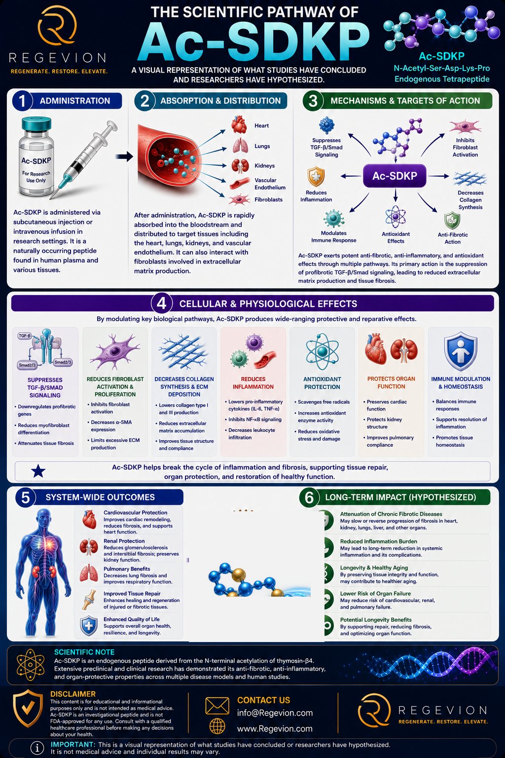
Ac-SDKP, formally N-acetyl-seryl-aspartyl-lysyl-proline, is a naturally occurring tetrapeptide with a distinctive position in human biology: it is both a signaling molecule and a metabolic substrate of angiotensin-converting enzyme (ACE). Its biology begins with thymosin β4 (Tβ4), a 43-amino-acid peptide; Ac-SDKP is the acetylated four-amino-acid sequence at Tβ4’s amino terminus, generated through sequential enzymatic processing involving meprin-α and prolyl oligopeptidase (POP). Once generated, it enters a tightly regulated metabolic cycle in which ACE is a major degradation route.¹
This creates a useful circuit: Tβ4 → enzymatic processing → Ac-SDKP → cellular signaling → ACE-mediated degradation. The ACE connection matters because ACE has two homologous catalytic domains — the N-terminal domain preferentially hydrolyzes Ac-SDKP, distinct from the better-known conversion of angiotensin I to angiotensin II.² ACE inhibition therefore does more than alter the renin-angiotensin system: it also reduces Ac-SDKP degradation and can substantially raise its circulating concentration. Ac-SDKP sits at an intersection between peptide metabolism, tissue remodeling, inflammation, and the renin-angiotensin system.
Molecular Mechanism of Action
Unlike many peptide drugs, Ac-SDKP has no single, universally established receptor that explains all of its effects. Experimental work has identified specific Ac-SDKP binding sites in cardiac fibroblasts, supporting receptor-mediated activity, but the receptor’s molecular identity hasn’t been definitively established³ — so its downstream biology is better described as a network of signaling effects than a fully resolved single-receptor pathway.
One of the most consistently investigated mechanisms involves transforming growth factor-beta (TGF-β), a major driver of tissue fibrosis: when TGF-β binds its receptors, Smad proteins become phosphorylated, form complexes, move into the nucleus, and increase expression of genes driving extracellular-matrix production and fibrotic remodeling. Ac-SDKP appears to oppose this architecture — experimental studies show reduced TGF-β-associated Smad2/3 phosphorylation in its presence, along with increased or redistributed Smad7 (an inhibitory Smad that brakes TGF-β receptor signaling), though the precise molecular sequence connecting Ac-SDKP to Smad regulation remains incompletely defined.⁴ The functional consequence is a shift away from persistent fibroblast activation and excessive matrix deposition:
Ac-SDKP exposure → suppression of profibrotic TGF-β/Smad signaling → less fibroblast activation and collagen production → reduced extracellular-matrix accumulation → attenuation of tissue fibrosis.
This has been demonstrated most strongly in experimental cardiovascular and renal models, not as a complete therapeutic mechanism established in humans.
Cellular Signaling Beyond TGF-β
Ac-SDKP appears to influence several additional cellular programs. In vascular and renal models it has been associated with suppressed inflammatory-cell infiltration and reduced cellular proliferation, implicating MAPK/ERK signaling, cell-cycle regulators, and fibroblast-behavior pathways. In human mesangial cells, it has been linked to increased expression of cell-cycle inhibitors p21 and p27, increased p53 activity, and reduced cyclin D1 — a potential explanation for reduced cellular proliferation.⁴
A further mechanistic layer involves endothelial-to-mesenchymal transition (EndMT), in which endothelial cells lose vascular-lining characteristics and acquire mesenchymal, fibroblast-like properties that can contribute to the population of activated fibroblasts. In diabetic kidney models, Ac-SDKP has been associated with restored fibroblast growth factor receptor (FGFR) signaling and increased expression of the antifibrotic let-7 microRNA family, with corresponding EndMT inhibition — a second mechanistic route by which it may limit fibrotic-cell accumulation.¹ These mechanisms shouldn’t be treated as separate effects: they converge on a common outcome of reducing the cellular programs that maintain chronic inflammation, proliferation, and matrix deposition.
Pharmacology: A Peptide Regulated by ACE
ACE isn’t simply an upstream receptor or signaling partner for Ac-SDKP — it’s also the metabolic enzyme that terminates its activity, preferentially using its N-terminal catalytic site to do so.² This creates a direct exposure relationship: more ACE activity → faster degradation → lower Ac-SDKP exposure; ACE inhibition → slower degradation → higher exposure. Human studies demonstrate this directly — captopril produced approximately a fivefold transient increase in plasma Ac-SDKP in healthy subjects, and chronic ACE-inhibitor treatment has been associated with substantially higher circulating concentrations.⁵ This is one reason Ac-SDKP has been proposed as a mediator of some ACE-inhibitor effects that blood-pressure reduction alone can’t explain. Importantly, Ac-SDKP is not itself an ACE inhibitor — it’s a substrate of the enzyme, which is a fundamentally different relationship from a conventional ACE-inhibiting drug.
Pharmacokinetics: What Happens to Ac-SDKP?
The pharmacokinetics of Ac-SDKP as an administered peptide are far less completely characterized than its endogenous metabolism. In circulation, it’s rapidly metabolized by plasma enzymes, with ACE the principal first step; an early human-plasma experiment measured an approximately 80-minute in-vitro half-life under the conditions used, and renal function also influences circulating concentration and clearance.⁶ Its circulating exposure therefore reflects a balance of generation → distribution → enzymatic degradation → renal handling — most visible in people on ACE inhibitors, where reduced degradation causes accumulation. There is currently insufficient human pharmacokinetic evidence to establish a standardized clinical exposure profile for exogenously administered Ac-SDKP, so experimental animal doses shouldn’t be translated directly into human dosing.
Dose, Exposure & Dose-Response
Dose-response relationships have primarily been studied preclinically. Dahl salt-sensitive rats have been studied at approximately 800 and 1,600 μg/kg/day, and renovascular-hypertension models at approximately 400 and 800 μg/kg/day via continuous subcutaneous infusion; higher exposure was sometimes associated with greater reductions in collagen accumulation or other injury markers.⁷ This illustrates dose → systemic exposure → tissue exposure → signaling intensity → biological response, but doesn’t establish an equivalent human dose. Ac-SDKP’s biology doesn’t support the assumption that more peptide always produces proportionally more benefit — cellular signaling can saturate, different endpoints can have different exposure-response relationships, and the concentration needed to affect a signaling pathway isn’t necessarily what’s needed for a whole-organ effect. The evidence supports exposure-dependent biological activity in experimental systems, but not a clinically validated human dose-response curve.
Does Body Weight Affect Dose?
There is no established human dosing framework showing Ac-SDKP should be dosed by body weight. Experimental studies commonly express administration as μg/kg/day — useful for controlling exposure relative to animal size — but that shouldn’t be read as evidence that human treatment requires weight-based dosing. No validated human pharmacokinetic model currently establishes how body weight alters distribution, clearance, or pharmacodynamic response enough to justify a specific weight-adjustment strategy — an important distinction between how researchers dose an animal experiment and how a clinically approved medicine would be dosed in humans.
Lower vs. Higher Exposure
Preclinical research suggests Ac-SDKP can influence tissue-remodeling pathways across a range of exposures. At lower exposures, effects may become detectable in specific molecular or inflammatory endpoints before larger structural changes appear; at higher exposures, some studies have observed stronger suppression of collagen accumulation and fibrosis-related pathology — in the Dahl salt-sensitive rat model, both 800 and 1,600 μg/kg/day reduced several measures of renal injury, while endpoints like albuminuria showed greater improvement at the higher exposure.⁷ This is why dose-response must be evaluated endpoint by endpoint: a biological pathway may respond before an anatomical outcome changes, and once a pathway nears maximal activation, increasing exposure may produce diminishing additional effect.
Connecting Molecular Mechanisms to Physiology
The most coherent interpretation of Ac-SDKP biology is a multilevel antifibrotic network: at the molecular level it interferes with profibrotic TGF-β/Smad signaling; at the cellular level this can reduce fibroblast proliferation, matrix production, inflammatory signaling, and potentially EndMT; at the tissue level, reduced collagen deposition limits pathological remodeling; at the organ level, experimental models show attenuated fibrosis in the heart and kidney; at the whole-body level, these effects can translate into preserved organ architecture and function in disease models. The important qualification: this chain is strongest in cellular and animal research — biologically plausible and repeatedly observed, but the complete causal sequence from administration to clinical benefit in humans hasn’t been established.
Combination & Pathway Interaction
Ac-SDKP’s biology intersects directly with the renin-angiotensin system, since ACE simultaneously participates in angiotensin metabolism and Ac-SDKP degradation — ACE inhibition changes both systems at once, reducing angiotensin II generation while increasing Ac-SDKP exposure. Experimental studies have shown combining Ac-SDKP with ACE inhibition can produce stronger antifibrotic effects than either alone in some models — in diabetic mice, combined treatment increased Ac-SDKP exposure and improved measures of renal fibrosis.⁸ This is mechanistically interesting rather than a treatment recommendation: it shows Ac-SDKP doesn’t operate in isolation, and its biological availability is intrinsically linked to ACE activity.
What Scientists Still Do Not Fully Understand
The central unresolved question is how Ac-SDKP initiates its intracellular signaling. Although binding sites have been identified experimentally, the definitive molecular identity of an Ac-SDKP receptor hasn’t been established,³ so the precise connection between surface recognition and downstream effects on TGF-β/Smad, MAPK, FGFR, microRNA, and cell-cycle pathways remains incomplete. Scientists also lack a complete human pharmacokinetic-pharmacodynamic model describing how circulating concentration translates into tissue exposure and biological response, and it remains unclear whether the antifibrotic effects seen across different organs arise from one conserved mechanism or from organ-specific signaling programs. Resolving these would help distinguish Ac-SDKP’s fundamental physiological role from effects specific to particular experimental conditions.
Why This Matters
Ac-SDKP is scientifically unusual because its biology connects peptide generation, enzymatic metabolism, intracellular signaling, inflammation, and tissue remodeling within one system — traceable from thymosin β4 processing, through ACE regulation, into cellular pathways like TGF-β/Smad and FGFR signaling, and ultimately into changes in fibroblast activity and extracellular-matrix deposition. That makes it more than a short peptide associated with reduced fibrosis: it’s a naturally occurring regulatory signal embedded within the body’s own enzymatic architecture. The most compelling science isn’t just that Ac-SDKP has been associated with reduced fibrosis in experimental models — it’s that researchers can trace how peptide availability changes cellular signaling, how those signals reshape tissue behavior, and how tissue-level remodeling can influence organ physiology. That mechanistic chain is the foundation of continued interest in Ac-SDKP.
References
- Ac-SDKP release from thymosin β4 by meprin-α and prolyl oligopeptidase; renal antifibrotic signaling (FGFR/let-7/EndMT). https://pubmed.ncbi.nlm.nih.gov/26962108/
- ACE N-terminal catalytic domain and Ac-SDKP hydrolysis. https://www.sciencedirect.com/science/article/pii/0968089696001046 ; https://pmc.ncbi.nlm.nih.gov/articles/PMC3982268/
- Characterization of Ac-SDKP receptor binding sites in cardiac fibroblasts. American Journal of Physiology-Heart and Circulatory Physiology. https://journals.physiology.org/doi/10.1152/ajpheart.00776.2006
- Ac-SDKP, TGF-β/Smad signaling, and cell-cycle regulation in renal antifibrotic mechanisms. https://pmc.ncbi.nlm.nih.gov/articles/PMC3995071/
- High plasma level of N-acetyl-seryl-aspartyl-lysyl-proline as a marker of ACE inhibition. https://pubmed.ncbi.nlm.nih.gov/9369248/ ; Azizi M, et al. J Clin Invest. 1996. https://www.jci.org/articles/view/118484
- Early human-plasma Ac-SDKP degradation kinetics and renal handling. https://pmc.ncbi.nlm.nih.gov/articles/PMC1137706/
- Dose-response studies in Dahl salt-sensitive and renovascular-hypertension rat models. https://pmc.ncbi.nlm.nih.gov/articles/PMC4658233/
- Combined Ac-SDKP and ACE-inhibitor treatment in diabetic mice (renal fibrosis). https://pmc.ncbi.nlm.nih.gov/articles/PMC4818806/