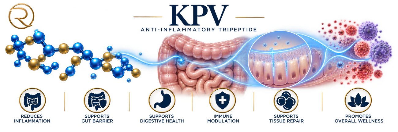
What the studies actually found
Researchers found that nanomolar concentrations of KPV suppress NF-κB and MAP kinase inflammatory signaling, and that it enters intestinal and immune cells via PepT1 — a transporter that ramps up specifically in inflamed gut tissue. Oral KPV reduced colitis severity in two chemically-induced mouse models.
READ THE RESEARCH →A separate German research team confirmed KPV's anti-inflammatory effect in two mouse colitis models, and — notably — showed it acts independently of the classical melanocortin receptors that its parent hormone, α-MSH, normally requires.
READ THE RESEARCH →Because PepT1 is elevated specifically at sites of gut inflammation, scientists engineered hyaluronic-acid-coated KPV nanoparticles designed to home in on damaged tissue. In a mouse model of ulcerative colitis, the targeted delivery reduced disease severity.
READ THE RESEARCH →As of 2026, the FDA's own review found no identified human clinical studies, human pharmacokinetic data, or human exposure data for KPV by any route. The cell and animal evidence is genuinely broad — skin, gut, airway, vascular models — but none of it is human proof yet.
READ THE RESEARCH →KPV is just three amino acids — lysine, proline, valine — clipped from the tail end of α-MSH, the hormone best known for controlling pigmentation. Somehow this tiny fragment kept the anti-inflammatory activity while losing the pigmentation effect entirely.
EXPLORE THE SCIENCE →KPV — Overview
What Is KPV?
KPV is a remarkably small peptide with a surprisingly broad scientific story. Its name comes from its three amino acids — Lysine–Proline–Valine — and corresponds to amino acids 11–13 at the C-terminal end of alpha-melanocyte-stimulating hormone (α-MSH), a naturally occurring 13-amino-acid peptide derived from the larger precursor protein proopiomelanocortin (POMC).
That origin matters because KPV wasn’t invented as an entirely new biological molecule. Scientists identified it as a small functional fragment within a much larger signaling peptide, then began asking a compelling question: could a three-amino-acid sequence preserve some of the useful biological activity of α-MSH without carrying all of its other effects? That question helped transform KPV from a small piece of a much larger hormone into a subject of independent research.
α-MSH is best known for its role in melanocortin biology, including pigmentation, but researchers discovered its influence extends well beyond skin color — the peptide participates in inflammation, immune regulation, fever, and tissue responses. Importantly, the anti-inflammatory activity of α-MSH was found to reside substantially in its C-terminal KPV sequence. KPV therefore represents an unusual category of biological molecule: a tiny peptide fragment that appears to retain meaningful biological activity from its much larger parent molecule.
Its simplicity is part of its scientific appeal — at only three amino acids long, KPV is substantially smaller than most biologically active peptides, making it interesting not only as a potential signaling molecule but as a model for understanding whether very short peptide sequences can influence complex biological processes such as inflammation. Unlike α-MSH, KPV doesn’t contain the complete structural motif required for conventional melanocortin-receptor binding, yet it retains much of the parent peptide’s anti-inflammatory activity without the pigment-producing effect associated with melanocortin signaling. The precise way KPV produces these effects remains an important area of investigation — and that distinction is central to why researchers continue to study it.
Scientific Discovery & Development
The story of KPV begins well before KPV itself became a research subject, with the discovery of the melanocortin system and the biological activity of melanocyte-stimulating hormones. By the late twentieth century, scientists were beginning to look more closely at the individual sequences within these hormones rather than treating the molecules as indivisible units. In a landmark structure-function study, researchers found that the C-terminal sequence containing Lys-Pro-Val could itself produce biological activity, demonstrating that α-MSH contained distinct functional regions capable of independently triggering biological responses.
The significance of KPV changed further when researchers began investigating inflammation. In 1989, James Lipton and Melante Hiltz reported that the C-terminal fragment of α-MSH — α-MSH(11–13), the Lys-Pro-Val sequence — could suppress inflammatory responses in experimental models, reducing chemically induced swelling in mouse tissue in a dose-related manner, suggesting this short fragment could carry an anti-inflammatory message of its own.¹ During the early 1990s, researchers continued examining the relationship between α-MSH fragments and inflammation, helping establish that the anti-inflammatory activity of melanocortin peptides could be separated from their pigmentary effects.
This created a new scientific opportunity: instead of using the entire α-MSH molecule, researchers could investigate its smallest active components. KPV became particularly interesting because it appeared to retain anti-inflammatory properties while lacking the structural features responsible for melanocortin-mediated pigmentation. By the 2000s, research had expanded into cellular inflammation, antimicrobial activity, intestinal biology, skin biology, and drug-delivery systems — the question had evolved from “Does KPV have biological activity?” to something more sophisticated: where does KPV act, how does it enter cells, and can its biological properties be used to influence inflammatory disease?
Why Researchers Are Studying KPV
The central scientific interest in KPV is inflammation. Inflammation isn’t inherently harmful — it’s one of the body’s fundamental defense and repair systems, and when tissues are damaged or threatened, immune and non-immune cells release signaling molecules that coordinate the response. The problem arises when inflammatory signaling becomes excessive, prolonged, or poorly controlled, so researchers are interested in molecules capable of reducing inappropriate inflammatory signaling without simply shutting down the immune system.
KPV is intriguing because laboratory research suggests it can influence important inflammatory pathways, including NF-κB and MAP kinase signaling — pathways near the center of many cellular responses to inflammatory stimuli, regulating the expression of numerous inflammatory mediators. This has led researchers to investigate KPV in several biological settings, particularly the intestinal tract and skin, where inflammation can profoundly affect tissue function.
The gut has been especially important to the KPV research story. Inflammatory bowel disease provides a useful experimental environment because intestinal inflammation involves epithelial cells, immune cells, cytokines, barrier function, and complex host-environment interactions. Researchers found that KPV could enter intestinal epithelial and immune cells through PepT1, a transporter normally involved in moving small peptides.² In experimental models, KPV reduced inflammatory signaling and improved measures of chemically induced colitis — a discovery significant not just because it demonstrated KPV could reduce inflammation, but because it suggested a potential biological route by which the peptide could reach cells involved in intestinal inflammation and influence their signaling from within.
Biological Foundation
To understand KPV, it helps to begin with its parent molecule. α-MSH is produced from POMC, a larger precursor protein processed into several biologically active peptides, and belongs to the melanocortin family, a signaling system involved in communication between the nervous, endocrine, immune, and peripheral tissues. The full α-MSH molecule can interact with melanocortin receptors distributed throughout the body, participating in diverse physiological functions.
KPV is different — it represents only the final three amino acids of α-MSH and doesn’t contain the complete receptor-binding architecture of the parent hormone, yet its biological behavior indicates a substantial portion of the anti-inflammatory “message” can survive in this much smaller sequence. This creates an interesting biological paradox: a molecule can be dramatically smaller than its parent hormone while retaining a specific portion of its biological activity.
Researchers have found that KPV can interact with the PepT1 transporter, particularly in intestinal epithelial and immune cells — a transporter for di- and tripeptides whose expression can increase in the colon under inflammatory conditions, potentially creating an important connection between intestinal inflammation and KPV uptake. Once inside cells, KPV has been associated with suppression of inflammatory signaling pathways, including NF-κB and MAPK pathways, and reductions in inflammatory cytokine production in experimental systems. The detailed molecular mechanism remains an active research question — the important question isn’t merely whether inflammation decreases in a laboratory model, but how a three-amino-acid peptide can influence the cellular machinery responsible for producing an inflammatory response. (The Science page goes deeper into this mechanism.)
What Researchers Are Investigating
Inflammatory bowel biology — the strongest area of KPV research, with the intestinal tract providing some of the clearest experimental evidence. In cellular and animal studies, researchers have observed reductions in inflammatory signaling and cytokine production following KPV exposure; in mouse models of chemically induced colitis, KPV administration reduced measures of intestinal inflammation and altered expression of inflammatory mediators.³ ⁴ This has led to investigation of KPV as a possible research tool for understanding inflammatory bowel disease and intestinal immune regulation, including whether it could influence the relationship between chronic inflammation and colitis-associated cancer in preclinical models — findings that remain preclinical and shouldn’t be interpreted as evidence KPV prevents or treats cancer in humans.
Skin biology — because inflammatory signaling is central to conditions involving irritated or damaged skin, researchers have investigated KPV in models involving keratinocytes, oxidative stress, and inflammatory responses. Recent laboratory work reported that KPV reduced inflammatory responses and cellular injury in human keratinocyte cultures exposed to fine particulate matter.⁵
Antimicrobial properties — experimental studies have reported activity against organisms including Staphylococcus aureus and Candida albicans, raising the possibility that melanocortin-derived peptides may participate in biological defense as well as inflammatory regulation. These findings are laboratory observations, not evidence of an established antimicrobial treatment.
Delivery — a three-amino-acid peptide may be attractive from a manufacturing and formulation perspective, but biological activity doesn’t automatically translate into effective delivery to human tissues. Researchers have investigated targeted oral delivery using nanoparticles and transdermal delivery systems, and experimental work has shown KPV can be incorporated into specialized delivery platforms designed to increase its exposure to intestinal or skin tissues.⁶ This illustrates an important principle in peptide research — discovering biological activity is only the beginning; scientists must also determine how a peptide reaches its target, how long it remains available, what dose produces a useful response, and whether the same effects occur safely in humans.
Potential Benefits & Biological Significance
KPV’s potential significance comes primarily from its relationship with inflammatory signaling. If the preclinical findings translate successfully to humans, KPV could eventually become interesting for conditions in which excessive inflammatory signaling contributes to tissue dysfunction — research has particularly focused on intestinal inflammation, inflammatory skin biology, tissue responses, and other immune-mediated processes.
The most scientifically compelling aspect isn’t simply that KPV appears anti-inflammatory — it’s that researchers have identified several pieces of a coherent biological story: a naturally occurring α-MSH fragment → preservation of anti-inflammatory activity → cellular uptake through PepT1 → modulation of inflammatory signaling → reduced inflammatory responses in experimental models. That chain gives KPV a stronger scientific rationale than a peptide whose proposed benefits are based primarily on anecdotal observations.
At the same time, the evidence has an important limitation: most of the research remains cellular or animal-based, and human clinical evidence specific to KPV is extremely limited — the absence of controlled human efficacy studies means potential benefits remain hypotheses rather than established medical outcomes. KPV is scientifically interesting because the biology is promising, not because its proposed applications have already been proven in people.
Current Research Stage & Future Outlook
KPV remains an investigational peptide and is not FDA approved for any medical indication. As of 2026, the most important recent regulatory development isn’t an approval but an FDA advisory process concerning compounding. On July 23, 2026, the FDA’s Pharmacy Compounding Advisory Committee reviewed KPV-related bulk drug substances for potential inclusion on the Section 503A Bulk Drug Substances List, specifically in connection with wound healing and inflammatory conditions. It’s worth distinguishing two separate things here: FDA staff’s own briefing document recommended against including KPV or KPV acetate on the list, citing no identified human exposure data or clinical studies and stating that important safety information remains lacking — but the advisory committee’s actual vote went the other way, reportedly recommending inclusion 8–6, with one abstention.⁷ That committee recommendation doesn’t make KPV an FDA-approved drug and doesn’t itself change KPV’s legal compounding status — advisory committee recommendations are non-binding, and the agency has stated that a final determination requires completion of its regulatory review process.
Scientifically, that leaves KPV at an important but early stage. The preclinical evidence has established a compelling biological hypothesis; the next major step is determining whether those mechanisms translate into meaningful, reproducible outcomes in humans, and whether an appropriate safety profile can be demonstrated.
Global Research Perspective
KPV research has developed through an international network of scientists rather than a single laboratory. Early work on α-MSH and its functional fragments involved researchers in the United States, Europe, and Japan. Later investigations into intestinal inflammation brought together research groups in the United States and China, including work involving Georgia State University, the Atlanta Veterans Affairs Medical Center, and collaborators at Fudan University. Research has also expanded into pharmaceutical science and drug delivery, with investigators at institutions including Auburn University, Tuskegee University, and California Health Sciences University examining approaches for transporting KPV across biological barriers such as the skin. What began as a question about a tiny fragment of a melanocortin hormone has developed into a multidisciplinary investigation involving peptide biology, immunology, gastroenterology, dermatology, molecular signaling, pharmaceutical delivery, and regulatory science.
Why This Matters
KPV is scientifically significant because it challenges a simple assumption about biological signaling — that meaningful biological effects always require large, complex molecules. This three-amino-acid fragment of α-MSH has preserved a remarkable portion of the parent peptide’s anti-inflammatory biology and has become the subject of research into how very small peptides can influence cellular inflammatory responses. Researchers have built a coherent preclinical case around KPV, particularly in intestinal and inflammatory biology, but the crucial transition from laboratory evidence to demonstrated human benefit has yet to occur — that gap is precisely why KPV deserves closer scientific attention. The Research page examines what the scientific literature has actually discovered over time; the Science page goes deeper into the molecular pathways, transport mechanisms, signaling systems, pharmacology, and biological responses that make KPV such an intriguing peptide.
References
- Hiltz ME, Lipton JM. Antiinflammatory activity of a COOH-terminal fragment of the neuropeptide α-MSH. The FASEB Journal. 1989. https://faseb.onlinelibrary.wiley.com/doi/10.1096/fasebj.3.11.2550304
- Dalmasso G, Charrier-Hisamuddin L, Nguyen HT, Yan Y, Sitaraman S, Merlin D. PepT1-mediated tripeptide KPV uptake reduces intestinal inflammation. Gastroenterology. 2008;134(1):166-178. https://pubmed.ncbi.nlm.nih.gov/18061177/
- Kannengiesser K, Maaser C, Heidemann J, et al. Melanocortin-derived tripeptide KPV has anti-inflammatory potential in murine models of inflammatory bowel disease. Inflammatory Bowel Diseases. 2008;14(3):324-331. https://pubmed.ncbi.nlm.nih.gov/18092346/
- Getting SJ, Schiöth HB, Perretti M. Dissection of the anti-inflammatory effect of the core and C-terminal (KPV) α-melanocyte-stimulating hormone peptides. 2003. https://pubmed.ncbi.nlm.nih.gov/12750433/
- Sung, et al. Lysine-Proline-Valine peptide mitigates fine dust-induced keratinocyte apoptosis. Tissue & Cell. 2025. https://pubmed.ncbi.nlm.nih.gov/40073467/
- Cheng J, et al. Inflammation-triggered self-immolative conjugates enable oral peptide delivery by overcoming gastrointestinal barriers. Science Advances. 2026. https://www.science.org/doi/10.1126/sciadv.aea2989
- FDA Briefing Document, Pharmacy Compounding Advisory Committee (PCAC) Meeting, July 2026 — KPV review (staff recommended against 503A inclusion, citing no identified human exposure data or clinical studies). https://www.fda.gov/media/193346/download ; committee vote (8-6, one abstention, in favor of inclusion) per meeting reporting.
KPV — The Research
How Was KPV Discovered?
The scientific story behind KPV begins not with KPV itself, but with the discovery of melanocyte-stimulating hormone (MSH) and the realization that this family of peptides could influence much more than pigmentation. In the early 1950s, Aaron B. Lerner and colleagues at Yale University were investigating the biological signals responsible for skin pigmentation, and their work helped establish MSH as a distinct pituitary hormone, with research published in 1954 and subsequent structural work identifying the melanotropins.
The next scientific shift was more important for KPV. Researchers began discovering that α-MSH was active in tissues involved in inflammation and immune regulation. By the late 1980s, scientists including James Lipton and Melante Hiltz were asking a more precise question: did the entire α-MSH molecule need to be present for its anti-inflammatory effects, or was a much smaller portion responsible? In 1989, Hiltz and Lipton reported that the C-terminal fragment of α-MSH, known as α-MSH(11–13), could reproduce anti-inflammatory activity.¹ That three-amino-acid sequence is KPV: lysine-proline-valine. The discovery was significant because it separated the anti-inflammatory activity from the larger hormone and suggested that a very small peptide could retain meaningful biological activity — creating a new research question: what exactly was KPV doing, and did it require the same receptors used by α-MSH?
From Hormone Fragment to Independent Research Target
During the 1990s and early 2000s, researchers increasingly treated KPV as more than simply a fragment of α-MSH, studying whether it could influence inflammatory signaling independently of the classical melanocortin receptors. A 2003 study by Simon Getting, Hans Björkman Schiöth, and Mauro Perretti was particularly important: researchers compared KPV with other α-MSH fragments in inflammatory models and found that KPV retained anti-inflammatory activity even when the classical melanocortin-receptor pathway didn’t adequately explain the response.² This helped establish that KPV has biological behavior distinct from the parent hormone.
Research in human keratinocytes added another piece of the puzzle. In 2004, investigators studying α-MSH, KPV, and related peptides found that these peptides could produce intracellular calcium responses in skin cells without the conventional cyclic-AMP response expected from classical melanocortin receptor activation.³ The findings reinforced the emerging view that KPV’s activity couldn’t simply be reduced to “miniature α-MSH.” The field was therefore moving from a simple observation — KPV reduces inflammation — toward a more interesting question: how does a three-amino-acid peptide produce that effect?
The Intestinal Research Breakthrough
One of the most influential developments came from research at Emory University School of Medicine led by Guillaume Dalmasso, Didier Merlin, and colleagues. In their 2008 Gastroenterology study, “PepT1-mediated tripeptide KPV uptake reduces intestinal inflammation,” researchers investigated whether intestinal cells could transport KPV into the cell and whether that uptake was connected to its anti-inflammatory effects.⁴ They found that KPV was transported through PepT1, a transporter capable of moving di- and tripeptides across cell membranes. In human intestinal epithelial cells and T cells, nanomolar concentrations of KPV reduced activation of inflammatory signaling pathways including NF-κB and MAP kinase pathways, while also reducing inflammatory cytokine production; oral KPV administration produced anti-inflammatory effects in two mouse models of colitis. The importance of this study wasn’t simply that inflammation decreased in mice — it provided a potential explanation for how KPV could enter intestinal cells and influence inflammatory signaling from within them, becoming one of the foundations for subsequent research into KPV and inflammatory bowel disease.
KPV and Inflammatory Bowel Disease
A closely related 2008 study by Katharina Kannengiesser and colleagues at the University of Münster examined KPV in two mouse models of intestinal inflammation.⁵ KPV-treated mice recovered body weight more effectively in a chemically induced colitis model, while examination of intestinal tissue showed fewer inflammatory infiltrates and lower myeloperoxidase activity. Importantly, KPV also retained activity — and even improved survival — in mice with a nonfunctional melanocortin-1 receptor. That observation mattered because it further weakened the idea that KPV’s effects were simply caused by conventional MC1R activation, supporting a developing scientific model in which KPV could act through mechanisms distinct from the receptor biology of full-length α-MSH.
Research Expands Beyond the Intestine
The research subsequently broadened into skin, airway, ocular, wound-healing, and vascular models. In airway epithelial cells, researchers found that KPV suppressed NF-κB signaling, IL-8, and eotaxin secretion, with evidence suggesting KPV could interfere with the movement of the NF-κB p65 subunit into the nucleus via its importin-α3 binding region.⁶ Research also explored whether KPV could influence tissue repair — in ocular research, α-MSH(11–13) was investigated for effects on corneal epithelial wound healing,⁷ while later studies examined delivery technologies designed to place KPV at sites where conventional peptide delivery is difficult.
This created an important secondary research field: delivery. Because KPV is a very small peptide, researchers began asking whether its therapeutic potential might depend as much on getting the molecule to the correct tissue as on the molecule’s intrinsic biological activity.
Delivery Became a Research Question
A 2017 study by Kasturi Pawar and colleagues at Auburn University examined whether KPV could cross human skin, in “Transdermal Iontophoretic Delivery of Lysine-Proline-Valine Peptide Across Microporated Human Skin,” published in the Journal of Pharmaceutical Sciences.⁸ Passive diffusion through intact skin was extremely limited; however, microneedles increased KPV penetration, and iontophoresis further increased delivery — the combination of microneedles and iontophoresis produced substantially greater penetration than passive diffusion alone. The significance was practical as well as scientific: KPV’s biological activity couldn’t automatically be translated into a topical therapy simply by putting the peptide on the skin. Delivery, stability, and tissue penetration became separate scientific problems that needed to be solved.
Newer Research Directions
More recent research continues to test whether KPV’s anti-inflammatory biology can be applied to additional conditions. In 2024, researchers incorporated KPV into a self-assembled nanoparticle system with rapamycin; in cell and mouse models of vascular calcification, the KPV-containing system reduced inflammatory activity and inhibited vascular calcification.⁹ This remains experimental nanomedicine research rather than evidence of a human treatment.
In 2025, researchers studying human keratinocytes found that KPV reduced inflammatory and oxidative effects caused by fine particulate matter exposure and improved cell viability under those experimental conditions.¹⁰
In 2026, research expanded into metabolic biology as well. A study using HepG2 liver cells reported that KPV reduced lipid accumulation and oxidative stress under experimentally induced steatosis, with effects involving ROS-dependent regulation of the PPARγ pathway.¹¹ These findings are cellular and shouldn’t be interpreted as evidence that KPV treats fatty liver disease in humans.
Another emerging direction is targeted oral delivery. Researchers have developed KPV-based delivery systems designed to protect the peptide from gastrointestinal degradation and release it preferentially in inflamed tissue. A 2026 study in Science Advances described an inflammation-triggered, self-immolative KPV conjugate: free KPV was largely degraded in simulated gastric fluid (roughly 9% survival at two hours), while the engineered conjugate achieved roughly 3.8-fold greater colonic accumulation at a 20-fold lower dose than free KPV in mouse colitis models, producing efficacy where free KPV showed none.¹² These studies illustrate where the field is heading — not simply asking whether KPV can influence inflammation, but whether researchers can control where, how long, and under what conditions KPV acts.
Human Research & Clinical Evidence
This is where the distinction between scientific interest and clinical evidence becomes especially important. KPV has been studied extensively in cells, animal models, and human-derived tissues, but that’s not the same as demonstrating efficacy in people. As of 2026, the FDA’s review found no identified clinical studies or human exposure data involving KPV drug products administered by any route, including no identified human pharmacokinetic or pharmacodynamic studies, and no clinical evidence establishing KPV’s effectiveness for wound healing or inflammatory conditions.¹³ This means the strongest evidence for KPV remains preclinical — the biological findings are substantial enough to justify continued investigation, but haven’t yet crossed the critical step of demonstrating clinical effectiveness in appropriately designed human trials.
What the Evidence Shows
Stronger evidence exists for the biological activity of KPV in laboratory systems and animal models. Across multiple experimental settings, researchers have repeatedly observed reductions in inflammatory signaling, including effects involving NF-κB and inflammatory cytokines; intestinal research has also provided evidence for PepT1-mediated cellular uptake.
Emerging evidence concerns tissue repair, skin inflammation, airway inflammation, vascular biology, metabolic signaling, and advanced delivery systems. These findings are scientifically interesting but remain largely cellular, ex vivo, or animal-based.
Early or experimental evidence includes applications such as fatty-liver biology, vascular calcification, and engineered oral delivery. These areas may expand the scientific understanding of KPV, but currently represent research questions rather than established therapeutic applications.
The most important limitation is therefore not a lack of interesting findings — it’s the absence of meaningful clinical evidence in humans.
Current Research Stage
KPV remains an investigational peptide and is not FDA approved. Its current position became particularly relevant in 2026, when the FDA formally evaluated KPV free base and KPV acetate for possible inclusion on the federal 503A Bulk Drug Substances List, in connection with proposed topical uses for wound healing and inflammatory conditions. FDA staff’s own July 2026 briefing document concluded that the available evidence was insufficient to establish effectiveness or human safety, and recommended against adding KPV or KPV acetate to the 503A Bulks List — the document specifically noted no identified information on human use of these substances and no published clinical studies of compounded KPV in humans.¹³
That staff recommendation, however, is distinct from what the advisory committee itself decided. Reporting on the July 23–24, 2026 Pharmacy Compounding Advisory Committee meeting indicates the committee’s actual vote went against the staff’s recommendation, voting 8–6 (with one abstention) in favor of recommending KPV for 503A inclusion — part of a broader pattern at that meeting, where the committee voted favorably on several peptides despite more cautious staff briefing positions. Either way, the advisory committee process is separate from FDA drug approval and doesn’t itself make KPV an approved therapy — a committee recommendation is non-binding, and the agency retains final regulatory authority.
The regulatory review is nevertheless scientifically useful because it provides a current independent assessment of the evidence: KPV has demonstrated pharmacological activity in preclinical models, but the clinical evidence needed to establish therapeutic effectiveness in humans is not yet available.
What Researchers Are Studying Now
The next stage of KPV research is increasingly focused on three questions. First, researchers are trying to determine the precise molecular targets responsible for KPV’s effects — evidence increasingly suggests classical melanocortin receptors don’t fully explain its activity, while pathways involving NF-κB, inflammatory cytokines, and PepT1 remain important areas of investigation. Second, researchers are working on delivery systems that can protect KPV, improve tissue penetration, and concentrate the peptide where inflammation is occurring. Third, researchers are testing whether the biological effects observed in cells and animals can eventually translate into meaningful human applications — that last question remains unanswered.
Future Scientific Potential
KPV remains scientifically interesting because an unusually small peptide has demonstrated a surprisingly broad range of biological activity across different experimental systems. The research has progressed from the original discovery that a three-amino-acid fragment of α-MSH could retain anti-inflammatory activity, to investigations of cellular transport, inflammatory signaling, tissue repair, and targeted delivery. What has been demonstrated is preclinical biological activity; what researchers are investigating is how to control and apply that activity therapeutically; what remains a possibility is whether those findings can ultimately be translated into safe and effective treatments for human inflammatory or tissue-related disorders. That transition — from compelling laboratory biology to demonstrated human benefit — is the next major scientific question for KPV.
Why This Matters
KPV is a good example of how peptide research can evolve from an unexpected biological observation into an entire field of investigation. Scientists first discovered that the smallest C-terminal fragment of α-MSH could preserve important anti-inflammatory activity; subsequent research showed that KPV could influence inflammatory signaling, enter cells through peptide transport systems, and produce measurable effects in several animal models. The research has also revealed an important limitation: biological activity doesn’t automatically equal clinical efficacy. Today, KPV remains scientifically interesting because the preclinical evidence provides a coherent foundation for continued investigation, while the absence of human clinical evidence leaves a clear and important question still to be answered.
References
- Hiltz ME, Lipton JM. Antiinflammatory activity of a COOH-terminal fragment of the neuropeptide α-MSH. The FASEB Journal. 1989. https://faseb.onlinelibrary.wiley.com/doi/10.1096/fasebj.3.11.2550304
- Getting SJ, Schiöth HB, Perretti M. Dissection of the anti-inflammatory effect of the core and C-terminal (KPV) α-melanocyte-stimulating hormone peptides. 2003. https://pubmed.ncbi.nlm.nih.gov/12750433/
- Elliott RJ, Szabo M, Wagner MJ, Kemp EH, MacNeil S, Haycock JW. Melanocortin receptor agonists induce Ca2+ signalling in human melanocytes and keratinocytes without cAMP elevation. Journal of Investigative Dermatology. 2004.
- Dalmasso G, Charrier-Hisamuddin L, Nguyen HT, Yan Y, Sitaraman S, Merlin D. PepT1-mediated tripeptide KPV uptake reduces intestinal inflammation. Gastroenterology. 2008;134(1):166-178. https://pubmed.ncbi.nlm.nih.gov/18061177/
- Kannengiesser K, Maaser C, Heidemann J, et al. Melanocortin-derived tripeptide KPV has anti-inflammatory potential in murine models of inflammatory bowel disease. Inflammatory Bowel Diseases. 2008;14(3):324-331. https://pubmed.ncbi.nlm.nih.gov/18092346/
- Land SC. Inhibition of cellular and systemic inflammation cues in human bronchial epithelial cells by melanocortin-related peptides: mechanism of KPV action and a role for MC3R agonists. International Journal of Physiology, Pathophysiology and Pharmacology. 2012;4(2):59-73. https://pubmed.ncbi.nlm.nih.gov/22837805/
- Effects of the COOH-terminal tripeptide α-MSH(11-13) on corneal epithelial wound healing: Role of nitric oxide. Experimental Eye Research. 2006.
- Pawar K, et al. Transdermal Iontophoretic Delivery of Lysine-Proline-Valine (KPV) Peptide Across Microporated Human Skin. Journal of Pharmaceutical Sciences. 2017.
- KPV and RAPA Self-Assembled into Carrier-Free Nanodrugs for Vascular Calcification Therapy. Advanced Healthcare Materials. 2024.
- Sung, et al. Lysine-Proline-Valine peptide mitigates fine dust-induced keratinocyte apoptosis. Tissue & Cell. 2025. https://pubmed.ncbi.nlm.nih.gov/40073467/
- Lysine-proline-valine peptide attenuates hepatic lipid accumulation through ROS-dependent regulation of the PPARγ pathway in HepG2 cells. Cytotechnology. 2026. https://link.springer.com/article/10.1007/s10616-026-00967-z
- Cheng J, et al. Inflammation-triggered self-immolative conjugates enable oral peptide delivery by overcoming gastrointestinal barriers. Science Advances. 2026. https://www.science.org/doi/10.1126/sciadv.aea2989
- FDA Briefing Document, Pharmacy Compounding Advisory Committee (PCAC) Meeting, July 2026 — KPV review. https://www.fda.gov/media/193346/download ; committee vote per meeting reporting.
KPV — The Science
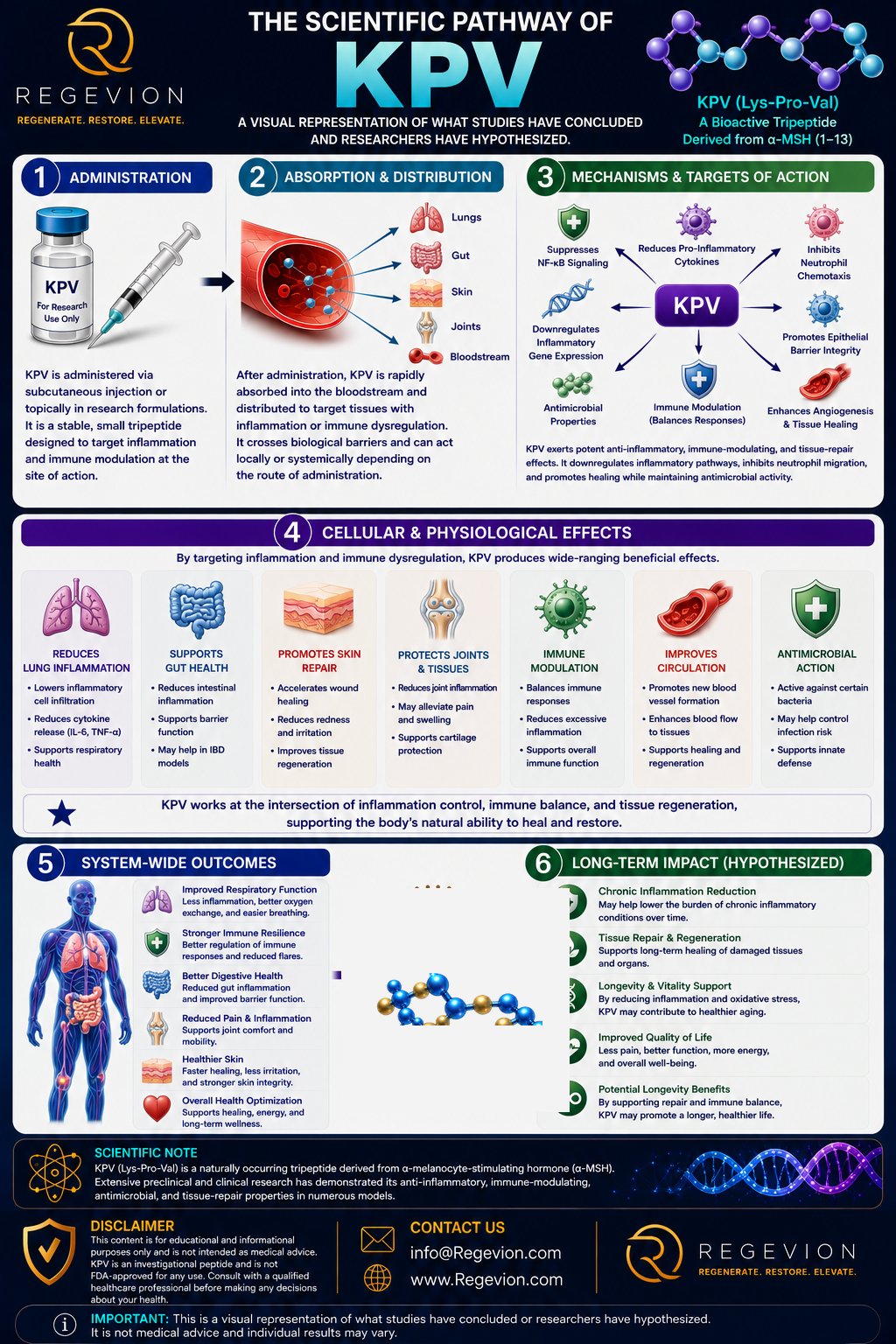
KPV, or Lys-Pro-Val, is a three-amino-acid peptide corresponding to residues 11–13 at the C-terminus of α-melanocyte-stimulating hormone (α-MSH). α-MSH is produced from the larger precursor protein proopiomelanocortin (POMC), which is processed in several tissues into biologically active melanocortin peptides.
The important scientific point is that KPV isn’t simply an arbitrary peptide fragment — it represents a biologically active sequence embedded within α-MSH. Early structure-function experiments showed that the Lys-Pro-Val sequence could retain biological activity independently of the complete α-MSH molecule. The broader melanocortin system communicates through five G-protein-coupled receptors: MC1R, MC2R, MC3R, MC4R, and MC5R, distributed across tissues including skin, immune cells, endocrine tissues, and the central nervous system. Full-length α-MSH can activate several melanocortin receptors and, through them, regulate processes ranging from pigmentation to immune activity.
KPV is unusual because its anti-inflammatory activity doesn’t appear to be explained simply by behaving as a miniature version of α-MSH at every melanocortin receptor. Experimental work points toward cellular uptake and direct intracellular regulation as important components of its activity — a distinction central to understanding KPV.
1. Molecular Mechanism of Action
The best-supported mechanistic model: KPV → cellular uptake → intracellular interaction → suppression of inflammatory signaling → reduced inflammatory mediator production.
One important route involves PepT1, a proton-coupled transporter normally responsible for moving small di- and tripeptides across intestinal epithelial cells; during intestinal inflammation, PepT1 expression can also increase in the colon. Research using human intestinal epithelial cells and immune-cell models found that KPV can be transported into cells through PepT1 — once inside, KPV suppressed activation of NF-κB and MAP kinase inflammatory signaling and reduced production of pro-inflammatory cytokines.¹ This creates a fundamentally different picture from a conventional peptide hormone acting only at a cell-surface receptor.
NF-κB functions as a major transcriptional regulator of inflammation. In an activated cell, inflammatory signals promote the release and nuclear movement of the NF-κB subunit p65/RelA; once inside the nucleus, NF-κB can activate genes involved in cytokine production, chemokine signaling, adhesion, and other components of the inflammatory response. KPV appears capable of interrupting this process. In human bronchial epithelial cells, KPV entered the nucleus and was associated with stabilization of IκBα, reduced nuclear translocation of p65/RelA, and inhibition of NF-κB-driven inflammatory signaling; the investigators also identified an interaction involving the importin-α3 binding region of p65/RelA, suggesting KPV may interfere directly with the machinery required to transport NF-κB into the nucleus.²
In simplified form: inflammatory stimulus → NF-κB activation → p65/RelA normally enters the nucleus → KPV interferes with this nuclear signaling step → reduced transcription of inflammatory mediators → lower cellular inflammatory signaling. This is one of the most distinctive mechanistic features of KPV.
2. Receptor Biology & Signaling
KPV’s relationship with the melanocortin receptor system is more complicated than simply labeling it an MC1R agonist. MC1R itself is a seven-transmembrane G-protein-coupled receptor associated primarily with Gs signaling and activation of adenylyl cyclase, which increases intracellular cyclic AMP (cAMP); in melanocytes, this pathway regulates transcriptional programs involving factors such as MITF and enzymes involved in melanin synthesis. α-MSH clearly uses MC1R to produce many of its classical effects. KPV, however, has produced anti-inflammatory responses in experimental systems where a straightforward MC1R → cAMP explanation doesn’t fully account for the observations.
In human keratinocyte experiments, KPV and related peptides produced intracellular calcium responses, but researchers didn’t observe the expected increase in cAMP that would establish a conventional MC1R-mediated mechanism.³ Similarly, airway research distinguished the mechanism of KPV from that of γ-MSH — γ-MSH suppressed inflammation through MC3R, whereas KPV acted through intracellular inhibition of NF-κB nuclear signaling.² The most defensible interpretation is that KPV can participate in melanocortin-related biology without its anti-inflammatory effects being reducible to classical melanocortin receptor activation. That distinction remains an important area of investigation.
3. Pharmacology
KPV has a very different pharmacological profile from a long-acting engineered therapeutic peptide. It’s only three amino acids long and structurally small enough to interact with peptide transport and intracellular systems in ways that larger peptide hormones may not; its small size also makes it susceptible to the normal processes that break down short peptides. Available research has focused predominantly on cellular and animal models rather than establishing a complete human pharmacokinetic/pharmacodynamic profile — robust human values for parameters such as receptor affinity, systemic half-life, bioavailability, volume of distribution, clearance, and exposure-response relationships haven’t been established to the standard expected for an approved drug. This limitation is scientifically important: a biological effect observed when KPV is placed directly into a cell culture system can’t automatically be translated into an equivalent systemic exposure in humans.
4. Pharmacokinetics: What Happens to KPV?
The most informative pharmacokinetic clue comes from its interaction with PepT1. PepT1 transports di- and tripeptides, and experimental work demonstrates KPV can use this transport system in intestinal epithelial and immune-cell models — providing a plausible mechanism by which KPV can enter cells rather than remaining exclusively outside them. However, this doesn’t establish that a particular oral, topical, or injected formulation will produce a predictable systemic concentration in humans. A 2026 engineered-conjugate study underscored this limitation directly: free KPV was largely degraded in simulated gastric conditions (roughly 9% survival at two hours), while a self-immolative, inflammation-responsive conjugate achieved substantially greater colonic accumulation at a much lower dose than free KPV in mouse colitis models.⁴ KPV’s short peptide structure means proteolytic stability, tissue exposure, route of administration, and local versus systemic delivery are likely important determinants of pharmacological activity. At present, the evidence is much stronger for understanding how KPV can enter cells and influence inflammatory signaling than for defining its complete human pharmacokinetic profile.
5. Dose, Exposure & Dose-Response
KPV research demonstrates that biological activity can be concentration-dependent, but the available evidence shouldn’t be interpreted as establishing a human therapeutic dose. In intestinal epithelial and immune-cell experiments, nanomolar concentrations of KPV inhibited NF-κB and MAP kinase inflammatory signaling;¹ in airway epithelial experiments, KPV also produced dose-dependent inhibition of NF-κB activity and inflammatory mediator release.² These experiments illustrate an important pharmacological principle: dose → cellular exposure → molecular interaction → signaling change → biological response. The relationship isn’t necessarily linear indefinitely — cellular targets can become saturated, intracellular transport can become limiting, and downstream signaling pathways can reach a functional ceiling — so increasing the concentration of KPV doesn’t automatically mean proportionally increasing its biological effect. There is currently insufficient human evidence to define clinically meaningful low-, moderate-, or high-exposure categories for KPV.
6. Does Body Weight Affect Dose?
There is currently no well-established human pharmacokinetic evidence demonstrating that KPV should be dosed according to body weight. Because human systemic exposure-response relationships haven’t been adequately characterized, there is also insufficient evidence to conclude that increasing or decreasing KPV exposure according to body size produces a predictable biological advantage — an important distinction between experimental concentration and validated clinical dosing.
7. Lower vs. Higher Exposure
Experimental systems provide evidence that KPV can suppress inflammatory signaling at biologically active concentrations, but they don’t establish a universal exposure threshold. At effective experimental concentrations, measurable changes include suppression of NF-κB activation, reduced inflammatory cytokine signaling, and decreased inflammatory mediator release. Higher concentrations may produce stronger inhibition within a particular experimental system, but this shouldn’t be interpreted as evidence that progressively higher exposure would produce proportionally greater effects in humans — the biological response is likely constrained by transporter availability, intracellular target interactions, signaling saturation, peptide stability, and tissue exposure.
8. Connecting Molecular Mechanisms to Physiological Effects
The mechanistic chain becomes particularly clear in experimental intestinal inflammation: KPV enters intestinal cells through PepT1 → intracellular inflammatory signaling is reduced → NF-κB and MAP kinase activity decrease → production of inflammatory cytokines and mediators falls → inflammatory cell recruitment and tissue injury can decrease → experimental intestinal inflammation is reduced. In mouse models of colitis, KPV treatment was associated with reduced inflammatory infiltration and lower tissue myeloperoxidase activity, providing a physiological bridge between the cellular signaling mechanism and tissue-level inflammatory outcomes.⁵ A similar mechanistic pattern appears in airway epithelial models, where inhibition of NF-κB signaling was accompanied by reduced IL-8, eotaxin, and MMP-9 activity.² The important scientific distinction is that these findings establish mechanistic plausibility and experimental activity, not proof that the same chain produces therapeutic benefit in humans.
9. Combination & Pathway Interaction
KPV operates at the intersection of several biological systems rather than through a single isolated pathway. Its relationship with the melanocortin system connects it to α-MSH biology, while its interaction with PepT1 provides a route into cells; once intracellular, its effects intersect with inflammatory signaling networks including NF-κB and MAP kinase pathways. This creates the possibility of pathway-level interactions in which KPV modifies the intensity of inflammatory signaling rather than acting as a single-target molecular switch. However, evidence for deliberate combination or “stacking” strategies is not sufficient to establish clinical compatibility, optimal combinations, or synergistic dosing — those questions require controlled pharmacological studies.
10. What Scientists Still Do Not Fully Understand
Several important questions remain open. The first is target identity: PepT1 clearly contributes to cellular uptake in intestinal models, but the complete intracellular molecular target or targets responsible for all of KPV’s anti-inflammatory effects haven’t been definitively established. The second is receptor independence — KPV is derived from α-MSH, yet its strongest anti-inflammatory mechanisms appear to involve intracellular processes that can’t simply be explained by conventional MC1R activation. The third is human pharmacokinetics: the field still lacks a sufficiently detailed human profile describing systemic exposure, tissue distribution, metabolic stability, and exposure-response relationships. Finally, recent literature continues to explore mechanisms beyond inflammation — for example, a 2026 study reported effects of KPV on oxidative stress and lipid-regulatory signaling in hepatocyte-derived (HepG2) cells, involving ROS and PPARγ pathways.⁶ These findings are mechanistically interesting but remain experimental and shouldn’t be treated as established human effects.
Why This Matters
KPV is scientifically interesting because its biology sits at the intersection of melanocortin signaling, peptide transport, intracellular trafficking, and inflammatory gene regulation. The emerging model isn’t simply that a small fragment of α-MSH activates a receptor — instead, KPV can enter relevant cells, interact with intracellular inflammatory machinery, and reduce signaling through pathways such as NF-κB. That creates a mechanistic bridge from peptide structure → cellular entry → molecular signaling → inflammatory gene regulation → tissue response. Understanding that chain is essential for interpreting the experimental evidence surrounding KPV — and for recognizing where the science is established, where it’s strongly supported, and where important questions remain unanswered.
References
- Dalmasso G, Charrier-Hisamuddin L, Nguyen HT, Yan Y, Sitaraman S, Merlin D. PepT1-mediated tripeptide KPV uptake reduces intestinal inflammation. Gastroenterology. 2008;134(1):166-178. https://pubmed.ncbi.nlm.nih.gov/18061177/
- Land SC. Inhibition of cellular and systemic inflammation cues in human bronchial epithelial cells by melanocortin-related peptides: mechanism of KPV action and a role for MC3R agonists. International Journal of Physiology, Pathophysiology and Pharmacology. 2012;4(2):59-73. https://pubmed.ncbi.nlm.nih.gov/22837805/
- Elliott RJ, Szabo M, Wagner MJ, Kemp EH, MacNeil S, Haycock JW. Melanocortin receptor agonists induce Ca2+ signalling in human melanocytes and keratinocytes without cAMP elevation. Journal of Investigative Dermatology. 2004.
- Cheng J, et al. Inflammation-triggered self-immolative conjugates enable oral peptide delivery by overcoming gastrointestinal barriers. Science Advances. 2026. https://www.science.org/doi/10.1126/sciadv.aea2989
- Kannengiesser K, Maaser C, Heidemann J, et al. Melanocortin-derived tripeptide KPV has anti-inflammatory potential in murine models of inflammatory bowel disease. Inflammatory Bowel Diseases. 2008;14(3):324-331. https://pubmed.ncbi.nlm.nih.gov/18092346/
- Lysine-proline-valine peptide attenuates hepatic lipid accumulation through ROS-dependent regulation of the PPARγ pathway in HepG2 cells. Cytotechnology. 2026. https://link.springer.com/article/10.1007/s10616-026-00967-z